Fractures - Foot
Disclaimer
These guidelines have been produced to guide clinical decision making for the medical, nursing and allied health staff of Perth Children’s Hospital. They are not strict protocols, and they do not replace the judgement of a senior clinician. Clinical common-sense should be applied at all times. These clinical guidelines should never be relied on as a substitute for proper assessment with respect to the particular circumstances of each case and the needs of each patient. Clinicians should also consider the local skill level available and their local area policies before following any guideline.
Read the full CAHS Clinical disclaimer
|
Mechanism of injury
- Crush injuries, stubbing of toes, kicking and tripping.
- Tarsal fractures are uncommon in isolation and are usually seen in crush injuries.
- Calcaneal fractures occur after a fall from a height landing on the heels. In these cases, compression fractures of the spine should also be considered.
Examination
- Localised pain, swelling and tenderness with reluctance to weight bear.
Imaging
- A foot X-ray should have dorsopalmar, oblique and lateral views.
- If there is midfoot tenderness, look carefully at the joint between the medial cuneiform and 2nd metatarsal base for a Lisfranc fracture as findings can be subtle.
- If the calcaneus is tender, dedicated axial calcaneal views must be requested as calcaneal fractures may not be visible on standard views.
- A Computerised Tomography (CT) scan may be indicated in talar fractures, intra-articular calcaneal fractures, severe crush injuries or suspicion of Lisfranc injury.
Specific management
Hindfoot fractures
Talar neck fractures
- Refer to the Orthopaedic team (high risk of avascular necrosis)
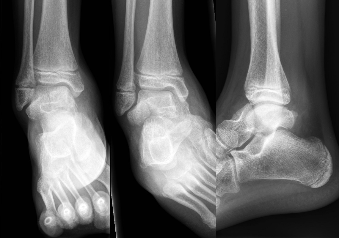
Intra-articular calcaneal fractures
- Refer to the Orthopaedic team
Minimally displaced talar and extra-articular calcaneal fractures
- Below knee backslab non weight bearing with crutches and follow up in the Orthopaedic Fracture clinic in 7 days.
Fracture of neck of talus
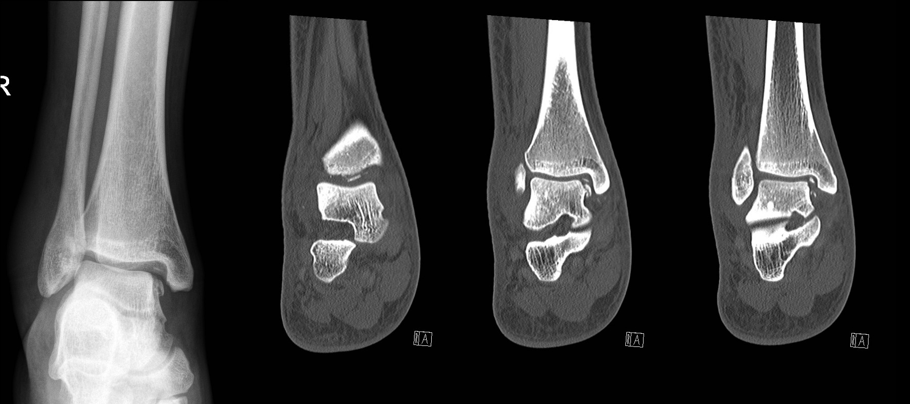
Talar dome fracture – looks simple on plain X-ray but CT shows multiple intra-articular bony fragments.
Midfoot fractures
Be wary of compartment syndrome in major midfoot fractures.
Avulsion fractures of the navicular or cuboid
Controlled Ankle Motion (CAM) boot weight bearing as tolerated with GP follow up in 7-10 days.
1
Tarsometatarsal fractures (Lisfranc disruption)
- Refer to the Orthopaedic team.
Metatarsal fractures
Undisplaced metatarsal fractures
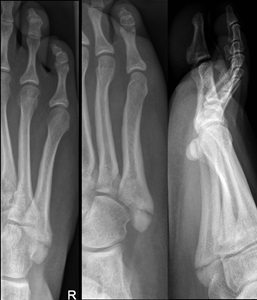
Minimally displaced oblique of the 5th metatarsal
Fractures of distal 2nd-4th metatarsals
- CAM boot weight bearing as tolerated with Fracture Clinic follow up in 1 week.1
Displaced metatarsal fractures
- Refer to the Orthopaedic Team.
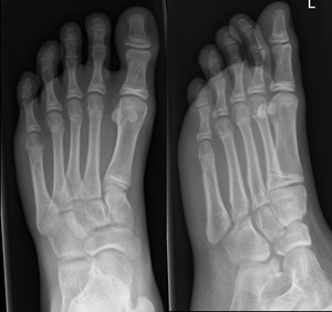
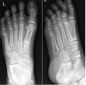
Avulsion fracture at the base of the fifth metatarsal (insertion of peroneus brevis)
Fracture of base of 5th metatarsal follow inversion injury
- CAM boot weight bearing as tolerated with Fracture Clinic follow up in 1 week.
- It is important to differentiate avulsion fractures of the base of the fifth metatarsal with Jones fractures which involve the 4th and 5th inter-metatarsal joint.
- These intra-articular fractures are managed in a below knee backslab non weight bearing with crutches and Fracture clinic follow-up in 1 week.
Phalangeal (Toe) Fractures
Open and intra-articular fractures
- Refer to the Orthopaedic team.
Big (1st) toe proximal phalanx fractures
- Darco Shoe non-weight bearing with crutches and follow up in Fracture Clinic in 1 week
Other phalangeal fractures (including distal phalangeal fractures of the big / 1st toe)

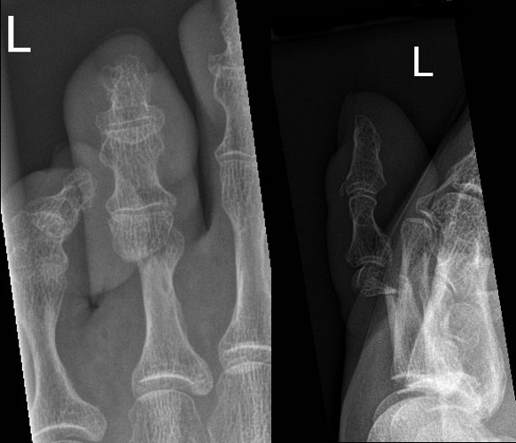
Angulated Salter-Harris II fracture of 5th proximal phalanx
Dorsally displaced transverse fracture of neck of 3rd proximal phalanx
- Consider reduction if significantly angulated.
- Buddy strap and Darco walking shoe or sturdy shoes with no specific follow up.
Phalangeal dislocations
- Relocate and buddy strap with no specific follow up.
References
- Bradman K, Stannage K, O’Brien S, Green, S et al. Randomised controlled trial comparing immobilisation in above-knee plaster of Paris to controlled ankle motion boots in undisplaced paediatric spiral tibial fractures. (2021) Emerg Med J 2021;0:1–7. doi:10.1136/emermed-2020-210299
- McRae R, Max Esser M, Practical Fracture Treatment Fifth Edition, Churchill Livingstone, 2008
- Rang M, Pring ME, Wenger DR. Kluwer W, Rang's Children's Fractures Fourth edition. Wolters Kluwer, 2018
| Endorsed by: |
Nurse Co-Director, Surgical Services |
Date: |
Feb 2024 |
This document can be made available in alternative formats on request for a person with a disability.