Chest trauma
Disclaimer
These guidelines have been produced to guide clinical decision making for the medical, nursing and allied health staff of Perth Children’s Hospital. They are not strict protocols, and they do not replace the judgement of a senior clinician. Clinical common-sense should be applied at all times. These clinical guidelines should never be relied on as a substitute for proper assessment with respect to the particular circumstances of each case and the needs of each patient. Clinicians should also consider the local skill level available and their local area policies before following any guideline.
Read the full CAHS clinical disclaimer
|
Aim
To guide Emergency Department (ED) staff with the assessment and management of children with major chest trauma.
Key Points1,2,5,6
- Note: TRAUMA CALL Triage Criteria - refer to Trauma - Serious Injury. The general surgery team should be involved early as part of a trauma call if indicated
- Thoracic injuries in children are rare and usually due to blunt trauma.
- The most common injuries are rib fractures, pulmonary contusions, pneumothoraces and haemothoraces.
- Management usually includes a combination of adequate oxygenation, analgesia, intravenous (IV) fluid / blood and a chest drain.
- Isolated major chest injuries are uncommon; they are usually associated with multisystem injury. Issues might result directly from chest injury or secondary to other injuries for example pulmonary aspiration secondary to vomiting.
- Initial stabilisation of a child with chest trauma requires identification of life-threatening injuries on primary survey and delayed life threats on secondary survey, following the catastrophic haemorrhage (cABCDE) approach.2,3
- Resuscitation and Primary Survey occur concurrently with treatment of problems as they are identified. The aim is to detect immediate life-threatening problems and to intervene as they are found.
- At Resuscitation: portable chest X-Ray (CXR) - unless signs of tension pneumothorax, address first.
- Once stabilised: consider thoracic Computerised Tomography (CT) scan after CXR if suspected major injury (rapid deceleration, high speed motor vehicle accident (MVA), or Glasgow Coma Score (GCS) < 14). CT Chest is unlikely to change management if the CXR and clinical examination is normal.8
- Many conditions worsen with time, especially pulmonary contusions. Continual clinical review is required (remember secondary survey).
Identify and resuscitate immediate life-threatening injuries concurrently. Treatment is prioritised over investigations in clear cut or unstable cases.
- Airway Obstruction / disruption
- Tension Pneumothorax
- Open Pneumothorax
- Massive Haemothorax
- Flail Chest
- Cardiac Tamponade
A: Airway: Airway obstruction or disruption
B: Breathing:
Tension Pneumothorax
- Clinical diagnosis
- Treatment precedes radiology in clear-cut or unstable cases
- Can occur in ventilated patients with a simple pneumothorax (undiagnosed or if no chest drain)
- Symptoms / signs:
- Tracheal deviation away from affected side (not always easily identifiable)
- Hypoxia
- Decreased air entry/movement, hyper-resonance/expansion (affected side)
- Tachycardia
- Hypotension (late sign)
|
Resuscitation
- Oxygen 15 L/min via nonrebreather mask
- Analgesia
- If patient is in cardiac arrest or peri-arrest: immediate needle decompression / needle thoracentesis (temporary measure). Refer to Appendix 1: Immediate Needle Decompression / Needle Thoracentesis Procedure for procedure details.
- For needle thoracostomy refer to Appendix 2: Needle Thoracostomy Procedure
|
Open Pneumothorax
- Penetrating chest wall injury with associated open pneumothorax
- Sucking air through the pleural space rather than the trachea during respiration.
- Symptoms / signs:
- Respiratory distress/failure
- Decreased air entry and chest wall movement
- Hyper-resonant to percussion
- Chest wall wound - air might be heard passing through with respiration.
|
Resuscitation
- Oxygen 15 L/min via nonrebreather mask.
- Analgesia
- In hospital
- Small wound: cover with occlusive dressing.
- Larger wound: suture/staple.
- Immediate formal intercostal catheter placed away from wound.
- Out of hospital
- 3-sided occlusive dressing (creating a one-way valve) – this is no longer recommended in a hospital setting.
- Consider use of 3-sided occlusive dressing in the Pre-hospital settings where there is a delay or chest drain insertion is not possible
- A bleeding or large wound could make this hard to apply
- An intercostal catheter should be placed away from wound when possible
|
Massive Haemothorax
- A hemithorax can hold up to 40% of a child’s blood volume
- Symptoms / signs:
- Signs of shock – tachycardia, hypotension, cold peripheries.
- Respiratory failure reduced air entry, chest wall movement and dullness to percussion.
Indications for urgent thoracotomy:1, 5
- Initial blood drainage >15-50 mL/kg of blood via chest drain.
- Continued bleeding > 2-3 mL/kg/hour continual.
- Significant residual haemothorax post-tube drainage.
|
Resuscitation
|
Flail Chest (chest wall instability or deformity)
- Paradoxical movement – the flail segment collapses with inspiration and bulge with expiration while the rest of the chest wall moves in the opposite way.
- Symptoms / signs:
- Respiratory distress.
- Possible decreased air entry due to associated pulmonary contusion and splinting.
- Possible subcutaneous emphysema.7
|
Resuscitation
- Oxygen 15 L/min via nonrebreather mask.
- Analgesia
- Stabilisation of flail segment – may be placed injured side down.
- Positive pressure ventilation.
- Improve ventilation.
- Intubation may be necessary to help with analgesic demands.
- Referral to Cardiothoracic surgeons for consideration of rib fixation.
|
C: Circulation
Analgesia should be initiated early and titrated to effect
- A structured approach will assist in identifying deteriorating patients with the aim of providing intervention early to prevent respiratory and cardiac failure resulting in cardiac or respiratory arrest. 2, 3, 5, 6
Cardiac Tamponade
- Bleeding into the pericardium secondary to blunt or penetrating injury
- Blood may originate from cardiac chamber, a great vessel or from the myocardium in the presence of a myocardial contusion
- This results in decreased filling of the heart and thus reduced stroke volume and shock
Symptoms /signs
- Respiratory distress
- Hypotension
- Narrow pulse pressure
- Distended neck veins
- Soft heart sounds
- Refractory shock
Investigations
- Electrocardiogram (ECG) – generally small complexes.
- Bedside ultrasound/eFAST for confirmation if patient is stable and an accredited clinician is available.
Cardiac arrest due to suspected traumatic cardiac tamponade: treat as per Traumatic Cardiac Arrest Guideline
|
Resuscitation
- Oxygen 15 L/min via nonrebreather mask
- ECG + ECG monitoring
- Fluid resuscitation (increase preload and minimise right ventricular collapse)
- Gold standard: Open surgical drainage in theatre
In severe shock / no surgical facilities available:
- Needle pericardiocentesis as a temporising measure.
- A long needle inserted at a left sub-xiphoid position, directed posteriorly at a 45° angle and towards the left shoulder.
- Best performed under ultrasound guidance where skills and equipment permit.
|
Severe haemodynamic instability, consider:
- Other bleeding source
- Tension pneumothorax
- Massive haemothorax
- Cardiac tamponade
- Tracheobronchial or mediastinal injury
- Pericardial injury
- Diaphragmatic injury
Consider need for Critical Bleeding protocol:
Indications for mechanical ventilation following chest trauma
- Ongoing respiratory distress / failure despite:
- Optimisation of oxygen delivery
- Adequate analgesia
- Chest drain insertion (when indicated)
- Closure of any open chest wounds
Indications for urgent surgical intervention7
- Massive haemorrhage
- Persistent haemorrhage
- Great vessel injury
- Pericardial tamponade
- Tracheobronchial injuries
- Oesophageal injury
- Diaphragmatic lacerations
- Open pneumothorax with major chest wall defect
- Penetrating chest trauma that crosses the mediastinum
ED Thoracotomy
Rarely indicated and only likely to be useful in a paediatric tertiary centre where the child can subsequently go directly to theatre with cardio-thoracic surgical facilities1,2,6.
Specific injuries1,2,3,5,6,7
Chest wall / Pulmonary
|
Rib Fractures
Rib fractures may be a marker of potential severe underlying injuries, especially if multiple or 1st rib fractures.
- Flail chest injuries are rare in children.
- Rib fractures in 0- to 3-year-old: consider non-accidental injury
|
Management
Oxygen supplementation in severe cases
|
Simple Pneumothorax
- Clinical signs: possible spectrum from no symptoms to decrease air entry and hyper resonance.
- Chest X-Ray: small pneumothorax may not be visible. Suspect if: increased radiolucency, deep sulcus sign.
|
Management
|
Pulmonary Contusion
- Can occur in the absence of rib fractures or other external signs.
- Chest X-Ray: increased pulmonary opacity, changes may evolve over time.
- CT scan: may reveal contusion not evident on initial Chest X-Ray
|
Management
- Oxygen Supplementation
- May require non-invasive or invasive ventilation.
- Analgesia
- Avoid excessive IV fluids.
|
Pulmonary Lacerations
- Penetrating injuries or associated with rib fractures.
- Cause haemothorax or pneumothorax and rarely, air embolism
|
Management
- Oxygen supplementation
- Management of concurrent injury
- Cardiothoracic surgical consultation
|
Tracheobronchial Injury
- Uncommon
- Clinical signs: subcutaneous emphysema, tension pneumothorax, haemoptysis, ongoing air leak
- Failure of lung expansion on chest X-Ray
|
Management
|
Mediastinal injury
- Most are rapidly fatal at the scene.
- Suspect based on mechanism of injury, physical signs (often absent) and chest X-Ray findings.
Chest X-Ray signs of aortic injury
- Widened mediastinum (mediastinum to chest radio >0.25)
- Loss or abnormal contour of aortic knob
- Depression of left mainstem bronchus
- Deviation of the trachea to the right
- Deviation of the oesophagus, nasogastric or orogastric tube to the right
- Left pleural cap.
- Left haemothorax
- Upper rib fractures
|
Management
- CT, angiography, aortogram or transoesophageal echocardiography
- Urgent cardiothoracic surgical referral
|
Cardiac Injuries
- Uncommon
- Pericardial tamponade / Myocardial contusion
- Tachycardia
- Elevation of jugular vein pressure (distended neck veins)
- Hypotension
- Arrest with pulseless electrical activity.
|
Management
- ECG
- Electrocardiography (echo)
- Urgent cardiothoracic surgical involvement
- Pericardiocentesis in arrested or unstable patient.
|
Oesophageal Trauma
- In penetrating trauma – high index of suspicion.
- Chest X-Ray – mediastinal air (early clue).
- With time (hours), an evolving sepsis with pleural effusion (usually left-sided) and mediastinitis ensue.
|
Management
- Gastrograffin study, Oesophagoscopy or both.
- Broad-spectrum antibiotics and urgent surgical review.
|
Diaphragmatic rupture
- Rare blunt injury
- More acknowledged now with penetrating injuries as occult injuries.
- Clinical signs: hypoxia.
- Chest X-Ray: show signs of herniated stomach or bowel.
- Laparotomy can confirm diagnosis.
|
Management
- CT scan – may miss small tears whilst magnetic resonance imaging has greater accuracy.
- Suspect occult diaphragmatic lacerations in penetrating trauma.
- Laparoscopy or thoracoscopy.
|
References and related external legislation, policies, and guidelines
- Cameron P, Browne GJ, Mitra B, et al., editors. Textbook of Paediatric Emergency Medicine. 4th ed. Elsevier; 2024.
- Advanced Paediatric Life Support (Australia & New Zealand): The Practical Approach. 7th ed. Wiley Blackwell; 2022.
- Eisenberg, M. Thoracic trauma in children: Initial stabilisation and evaluation. [Internet]. UpToDate. 2023. [Cited 9 Jun 2026]/ Available from: Thoracic trauma in children: Initial stabilization and evaluation - UpToDate
- Quinn N, Ward G, Ong C, Krieser D, Melvin R, Makhijani A, et al., Mid-Arm Point in PAEDiatrics (MAPPAED): An effective procedural aid for safe pleural decompression in trauma. Emerg Med Australas. [Internet]. 2023. [Cited 6 Jun 2026]. 35(3):412-419. Available from: https://doi.org/10.1111/1742-6723.14141
- Starship Health. Chest Injury. [Internet] Starship Health; 2023. [Cited 9 Jun 2026]. Available from: Chest Injury
- Royal Children’s Hospital. Chest Trauma Clinical Practice Guideline. [Internet]. 2023. [Cited 9 Jun 2026]. Available from: Trauma Service : Chest injury
- Starship Child Health. Chest injury - thoracocentesis, thoracostomy and intercostal catheter insertion. [Internet]. 2025. [Cited 9 Jun 2026]. Available from: https://www.starship.org.nz/guidelines/chest-injury-thoracocentesis-thoracostomy-and-intercostal-catheter-insertion/
- Royal College of Radiologists. Major paediatric trauma radiology guidance [Internet]. Royal College of Radiologists. 2024. [Updated Oct 2024; Cited 9 Jun 2026]. Available from: https://www.rcr.ac.uk/media/1q2eroqj/rcr-major-paediatric-trauma-radiology-2024_.pdf
Appendix 1: Immediate Needle Decompression / Needle Thoracentesis Procedure
- 16 G/14 G cannula + 10 mL syringe with 2 mL sodium chloride 0.9%.
- Once air is aspirated, advance cannula only. Remove the needle and syringe.
- Advance cannula to second intercostal space, mid-clavicular line OR
- Between the anterior and mid-axillary line on ipsilateral side (provides a larger zone of safety than the anterior approach).
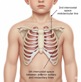
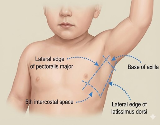
Image outlines landmarks for chest needle decompression
Appendix 2: Needle Thoracostomy Procedure
- Always follows needle decompression.
- Incision into 4th or 5th intercostal space between anterior and mid-axillary line
- Followed by a finger sweep (mosquito or artery forceps for blunt dissection to pleura
- Follow Mid-Arm Point in PAEDiatrics technique (MAPPAED) – the MAPPAED rule provides an age-based adjustment for safe pleural decompression site identification in children4.
- Mid-Arm Point (MAP): Halfway point between acromion and olecranon, transposed to the chest wall
- <4 years, use MAP
- ≥ 4 years, use MAP plus go one intercostal space above
- Place a formal Intercostal Catheter
| Endorsed by: |
Nurse Co-director, Surgical Services |
Date: |
Jul 2026 |
This document can be made available in alternative formats on request for a person with a disability.