Fractures - Forearm
Disclaimer
These guidelines have been produced to guide clinical decision making for the medical, nursing and allied health staff of Perth Children’s Hospital. They are not strict protocols, and they do not replace the judgement of a senior clinician. Clinical common-sense should be applied at all times. These clinical guidelines should never be relied on as a substitute for proper assessment with respect to the particular circumstances of each case and the needs of each patient. Clinicians should also consider the local skill level available and their local area policies before following any guideline.
Read the full CAHS clinical disclaimer
|
See Fractures – Overview for general assessment and management
Mechanism of Injury
- Falls onto an outstretched hand (FOOSH) with forward momentum.
- Direct blows to the forearm account for a smaller proportion of radius and ulna shaft fractures.
Examination
- There is usually swelling, tenderness and decreased range of movement, especially pronation and supination.
- There will be obvious deformity with displaced fractures.
- Remember to always examine the elbow and wrist joints.
Imaging
- A forearm X-ray should have AP (anterior-posterior) and lateral views and includes true AP and lateral views of the wrist and elbow joints.
- Assess the radiocapitellar line and radio-ulnar joint. See Fractures - Elbow.
Specific management
Midshaft radius and ulna
- Midshaft fractures may be greenstick, complete or bowed (plastic deformity, usually < 10 years old).
Fractures with minimal displacement, < 10 degrees volar angulation and < 20 degrees dorsal angulation
- Above elbow backslab with Orthopaedic Fracture clinic follow up in 7-10 days.
- Consider gentle moulding of the backslab under Nitrous Oxide / Oxygen sedation to improve angulation – discuss with Emergency Department (ED) senior doctor.
Fractures with clinical deformity, > 10 degrees volar angulation, > 20 degrees dorsal angulation, or any displacement
- Discuss with Orthopaedic team for possible reduction.
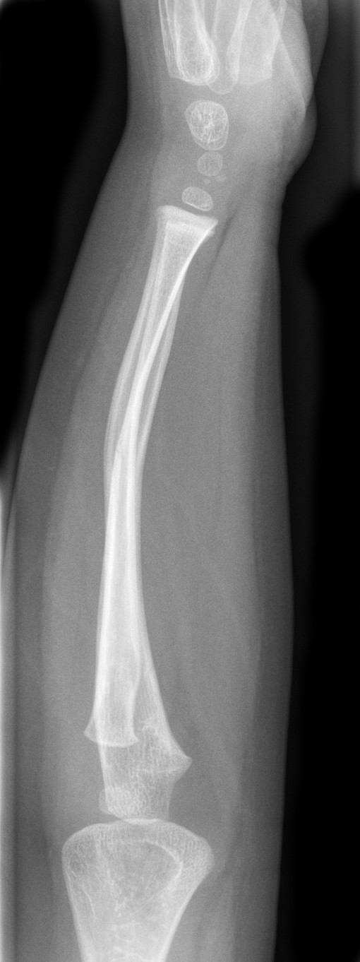
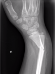
Plastic deformity of radius and ulna with volar tilt (left)
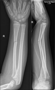
Greenstick fracture of radius shaft with 20 degrees of dorsal angulation and minimally displaced ulna fracture (right)
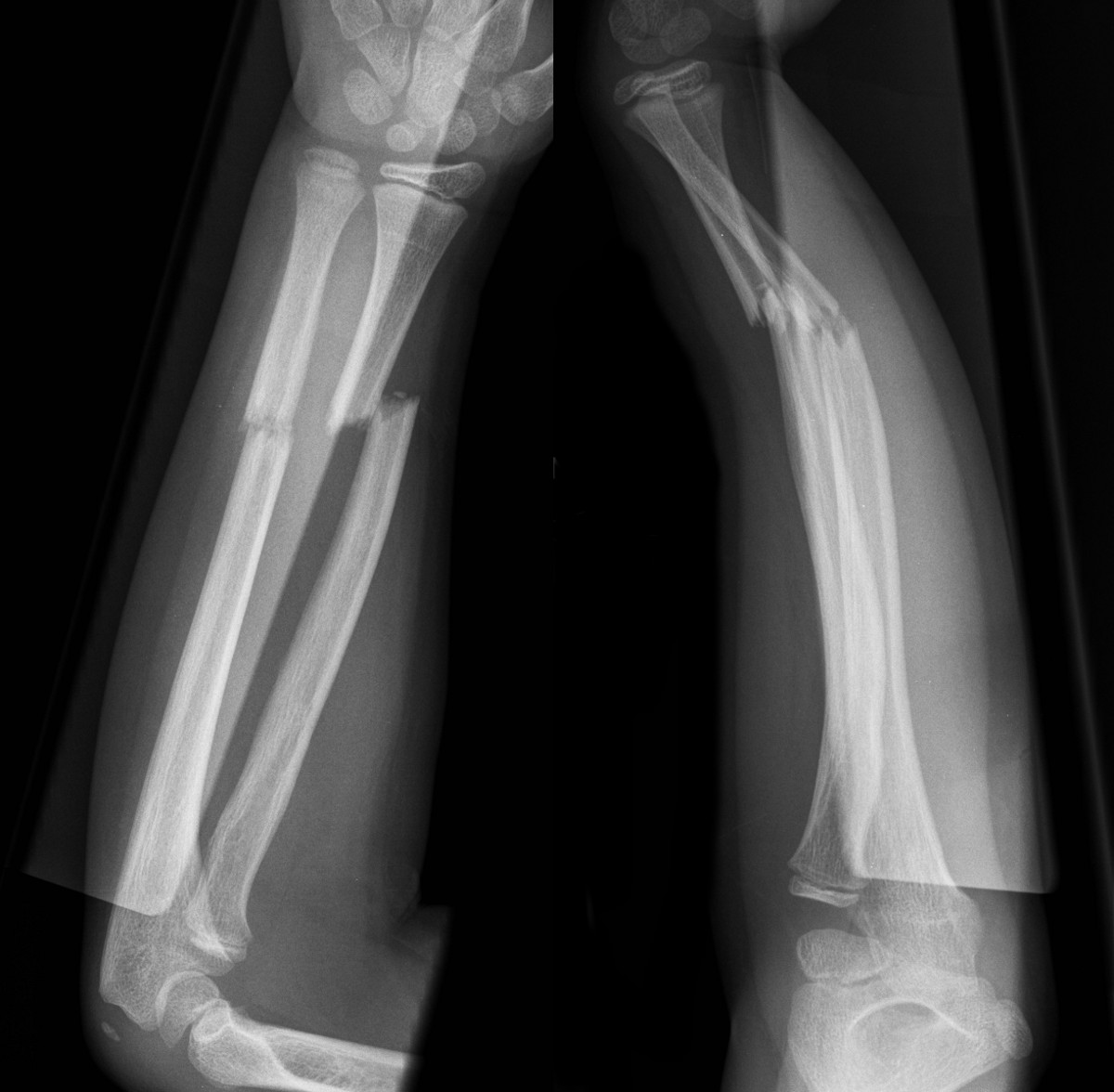
Transverse fractures of radius and ulna with complete displacement of radius and significant dorsal angulation (above)
Monteggia Fracture-Dislocation
- Proximal or mid-third ulna fracture, with an associated dislocation of the radial head.
- Occurs from 2 years of age to puberty.
- Isolated ulna shaft fractures are rare, always look for a radial head dislocation.
- There may be elbow swelling and pain in addition to an obvious forearm (ulna) fracture.
- Always assess the radiocapitellar line on lateral elbow radiographs (see Fractures - Elbow) as radial head dislocations are often missed.
- Isolated radial head dislocation never occurs. Always look for an associated ulna fracture which may be a subtle plastic deformity.
- All Monteggia fracture-dislocations should be referred to the Orthopaedic team for reduction. Apply an above elbow resting backslab.
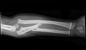
Monteggia fracture dislocation - ulna shaft fracture with radial head dislocation
Galeazzi fracture dislocation
- Radial shaft fracture (usually distal third) with distal radioulnar joint disruption
- Clinical distal ulnar prominence with joint instability may be present
- Galleazzi fractures should be referred to the Orthopaedic team urgently for reduction. Apply an above elbow resting backslab.
Galeazzi fracture dislocation
References
- McRae R, Max Esser M, Practical Fracture Treatment Fifth Edition, Churchill Livingstone, 2008
- Rang M, Pring ME, Wenger DR. Kluwer W, Rang's Children's Fractures Fourth edition. Wolters Kluwer, 2018
| Endorsed by: |
Co-director Surgical Services (Nursing) |
Date: |
Mar 2024 |
This document can be made available in alternative formats on request for people with disability.